Showing posts with label estrogen. Show all posts

Tuesday, January 23, 2024

My wish list for 2024: Hormonal inputs into perception of human skin color by men and women


Subjects identify the face on the left as female and the face on the right as male. The only difference is the lightness of the skin. Richard Russell, Sinha Laboratory for Vision Research, MIT.

--------------------------------------------------------------------------------


“The fair sex” is paler than men, who conversely are ruddier and browner than women. This sex difference seems to play a role in gender recognition and in relations between men and women, particularly in female response to darker male skin.


Women are universally the fair sex. They are paler than men, who conversely are ruddier and browner (Frost, 2010; Frost, 2023; van den Berghe and Frost, 1986). This sex difference is due to the differing ways the skin’s pigments—melanin, hemoglobin, carotene—interact with the sex hormones, either androgens in men or estrogens in women. A hormonal cause has been shown by studies of normal, castrated, and ovariectomized individuals, by studies of skin reflectance at puberty, and by studies of digit ratios (Edwards and Duntley, 1939; Edwards et al., 1941; Edwards and Duntley, 1949; Frost, 1988; van den Berghe and Frost, 1986; Manning et al., 2004).


Gender recognition


This sex difference is used subconsciously to recognize male and female faces (Frost, 2011; Russell, 2003; Russell, 2009; Russell, 2010; Russell et al., 2006; Semin et al., 2018).

Specifically, gender is identified from two aspects of facial color:


·         hue (men are ruddier and browner)

·         brightness (facial skin is lighter in women and contrasts more with the darker lip/eye area).


Hue provides the observer with a fast channel for gender recognition. If a face is too far away or the lighting too dim, the observer will switch to the slower but more accurate channel of brightness (Dupuis-Roy et al., 2009; Dupuis-Roy et al., 2019; Jones et al., 2015; Nestor and Tarr, 2008a; Nestor and Tarr 2008b; Tarr et al. 2001; Tarr, Rossion, and Doerschner, 2002). We thus perceive skin color through the lens of a mental algorithm that arose for gender recognition. This algorithm may explain why lighter skin seems more feminine and darker skin more masculine (Semin et al., 2018).


Male-female relations


The differing complexions of men and women play a role not only in gender recognition but also in relations between men and women. In particular, it seems to play a role in attraction by women to men.


In one study, women were asked to optimize the attractiveness of facial pictures by varying the skin's darkness and ruddiness. They made the male faces darker and ruddier than the female faces (Carrito et al., 2016). In another study, women were asked to rate different levels of male ruddiness. They associated high levels with aggression, medium levels with dominance, and low levels with attractiveness. Unlike the participants of the first study, they may have understood the term “attractive” in an aesthetic or even feminine sense (Stephen et al., 2012).


Female attraction to darker, ruddier male skin seems to be mediated by the level of estrogen in brain tissues. This estrogenic effect is shown by two studies of women at different phases of their menstrual cycle and by a study of preschool children:


·         Women were shown pairs of facial pictures that differed slightly in the lightness of the skin, and they were asked to choose the most pleasing one. When male faces were shown, the darker one was more strongly preferred by those women who were in the first two-thirds of their menstrual cycle than by those in the last third. During the first two-thirds of the cycle, the level of estrogen is high in relation to the level of progesterone (which acts as an anti-estrogen). During the last third, the ratio is reversed: the level of estrogen is low in relation to the level of progesterone. There was no cyclical effect among women judging female faces or taking oral contraceptives (Frost, 1994).


·         Women had their brain activity measured by MRI while viewing pictures of male faces. Their brains showed a stronger response to masculinized male faces than to feminized ones, and the strength of their response correlated with the level of estrogen across the menstrual cycle. In a personal communication, the lead author stated that the faces had been masculinized by making them darker and more robust in shape (Rupp et al., 2009).


·         Preschool boys and girls were presented with two dolls that differed slightly in skin color and asked to choose the “nicer” one. Their choices were recorded, as were measurements of their body mass index and their subcutaneous fat. Doll choice did not differ by sex. But it did differ by adiposity. Among children less than three years old, those who chose the darker doll had significantly more body fat than those who chose the lighter doll. In that age range, estrogen is produced mostly in the fatty tissues, which contain an enzyme (aromatase) that converts an androgen (androstenedione) into an estrogen (estrone) (Baird, 1976; Frost, 1989).


The doll on the right is slightly darker and ruddier than the one on the left. Among children below three years of age, those who chose the darker doll had significantly more body fat than those who chose the lighter doll. At such ages, estrogen is produced mainly in the body’s fatty tissues.

---------------------------------------------------------------------------------------------


In other doll studies, boys and girls have similar preferences up to six years of age (Renninger and Williams, 1966; Williams and Roberson, 1967; Williams and Rousseau, 1971). At older ages, male and female preferences begin to diverge. When a group of American children, 3 to 8 years of age, were presented with a white-faced puppet and a brown-faced one, the latter puppet was more often chosen by girls than by boys, this finding being as true for Euro-American children as for African American children (Asher and Allen, 1969).


There are fewer controlled studies of male response to lighter female skin. It has been argued that the lighter skin of women mimics that of infants, whose pinkish color is especially noticeable in darker-skinned populations and, apparently, in other primate species. It seems to identify the primate infant as a vulnerable being in need of protection (Alley, 1980; Booth, 1962; Jay, 1962).


In our species, the adult female may have evolved a lighter complexion as a means to tap into the same behavioral response, the aim being not so much to increase male sexual arousal as to reduce male aggressiveness and stimulate feelings of care (Frost, 2010, p. 131-136; Frost, 2023; Guthrie, 1970).


Proposed study


First research aim: expand on Rupp et al. (2009) by using brain MRI to measure how women respond to male facial hue and luminosity in relation to the levels of estrogen and progesterone across the menstrual cycle. Male facial photos would be altered to produce different degrees of brownness, redness, and brightness.


Second research aim: repeat the doll study of Frost (1989) with direct measures of estrogen and androgen levels in preschool children. This may be difficult, given the low hormonal levels of early childhood (Baird, 1976; Klein et al., 1994).


References


Alley, T. R. (1980). Infantile colouration as an elicitor of caretaking behaviour in Old World primates. Primates 21(3): 416-429. https://doi.org/10.1007/BF02390470


Asher, S.R. and Allen, V.L. (1969). Racial preference and social comparison processes. Journal of Social Issues 25(1): 157-166. https://doi.org/10.1111/j.1540-4560.1969.tb02584.x    


Baird, D.T. (1976). Oestrogens in clinical practice. In: J.A. Loraine and E. Trevor Bell (eds.) Hormone assays and their clinical application (p. 408). Edinburgh: Churchill Livingstone.


Booth, C. (1962). Some observations on behavior of Cercopithecus monkeys. Annals of the New York Academy of Sciences 102(2): 477-487. https://doi.org/10.1111/j.1749-6632.1962.tb13654.x   


Bruce, V., and Langton, S. (1994). The use of pigmentation and shading information in recognising the sex and identities of faces. Perception 23(7): 803-822. http://dx.doi.org/10.1068/p230803   


Carrito, M.L., dos Santos, I.M.B., Lefevre, C.E., Whitehead, R.D., da Silva, C.F., and Perrett, D.I. (2016). The role of sexually dimorphic skin colour and shape in attractiveness of male faces. Evolution and Human Behavior 37(2): 125-133. https://doi.org/10.1016/j.evolhumbehav.2015.09.006    


Dupuis-Roy, N., Faghel-Soubeyrand, S., and Gosselin, F. (2019). Time course of the use of chromatic and achromatic facial information for sex categorization. Vision Research 157: 36-43. https://doi.org/10.1016/j.visres.2018.08.004   


Dupuis-Roy, N., Fortin, I., Fiset, D., and Gosselin, F. (2009). Uncovering gender discrimination cues in a realistic setting. Journal of Vision 9(2): 10, 1-8. https://doi.org/10.1167/9.2.10   


Edwards, E.A., and Duntley, S.Q. (1939). The pigments and color of living human skin. American Journal of Anatomy 65(1): 1-33. https://doi.org/10.1002/aja.1000650102   


Edwards, E.A., and Duntley, S.Q. (1949). Cutaneous vascular changes in women in reference to the menstrual cycle and ovariectomy. American Journal of Obstetrics & Gynecology 57(3): 501-509. https://doi.org/10.1016/0002-9378(49)90235-5   


Edwards, E.A., Hamilton, J.B., Duntley, S.Q., and Hubert, G. (1941). Cutaneous vascular and pigmentary changes in castrate and eunuchoid men. Endocrinology 28(1): 119-128. https://doi.org/10.1210/endo-28-1-119   


Frost, P. (1988). Human skin color: A possible relationship between its sexual dimorphism and its social perception. Perspectives in Biology and Medicine 32(1): 38-58. https://doi.org/10.1353/pbm.1988.0010


Frost, P. (1989). Human skin color: the sexual differentiation of its social perception. Mankind Quarterly 30: 3-16. http://doi.org/10.46469/mq.1989.30.1.1   


Frost, P. (1994). Preference for darker faces in photographs at different phases of the menstrual cycle: Preliminary assessment of evidence for a hormonal relationship. Perceptual and Motor Skills 79(1): 507-14. https://doi.org/10.2466/pms.1994.79.1.507   


Frost, P. (2010). Femmes claires, hommes foncés. Les racines oubliées du colorisme. Quebec City: Les Presses de l'Université Laval, 202 p. https://www.pulaval.com/livres/femmes-claires-hommes-fonces-les-racines-oubliees-du-colorisme    


Frost, P. (2011). Hue and luminosity of human skin: a visual cue for gender recognition and other mental tasks. Human Ethology Bulletin 26(2): 25-34. https://www.researchgate.net/publication/256296588_Hue_and_luminosity_of_human_skin_a_visual_cue_for_gender_recognition_and_other_mental_tasks    


Frost, P. (2023). The original meaning of skin color. Aporia Magazine, February 7.


Guthrie, R.D. (1970). Evolution of human threat display organs. In T. Dobzhansky, M.K. Hecht, and W.C. Steere (Eds.) Evolutionary Biology 4: 257-302. New York: Appleton-Century Crofts.


Hill, H., V. Bruce, and Akamatsu, S. (1995). Perceiving the sex and race of faces: The role of shape and colour. Proceedings of the Royal Society B: Biological Sciences 261(1362): 367-373. https://doi.org/10.1098/rspb.1995.0161   


Hill, R., and Barton, R. (2005). Red enhances human performance in contests. Nature 435: 293. https://doi.org/10.1038/435293a   


Jay, P.C. (1962). Aspects of maternal behavior among langurs. Annals of the New York Academy of Sciences 102(2): 468-476. https://doi.org/10.1111/j.1749-6632.1962.tb13653.x


Jones, A.L., Russell, R., and Ward, R. (2015). Cosmetics alter biologically-based factors of beauty: evidence from facial contrast. Evolutionary Psychology 13(1): https://doi.org/10.1177%2F147470491501300113    


Klein, K.O., Baron, J., Colli, M.J., McDonnell, D.P., and Cutler, G.B. Jr. (1994). Estrogen levels in childhood determined by an ultrasensitive recombinant cell bioassay. Journal of Clinical Investigation 94(6): 2475-2480. https://doi.org/10.1172/JCI117616


Manning, J.T., Bundred, P.E., and Mather, F.M. (2004). Second to fourth digit ratio, sexual selection, and skin colour. Evolution and Human Behavior 25(1): 38-50. https://doi.org/10.1016/s1090-5138(03)00082-5   


Nestor, A., and Tarr, M.J. (2008a). The segmental structure of faces and its use in gender recognition. Journal of Vision 8(7): 7, 1-12, https://doi.org/10.1167/8.7.7   


Nestor, A., and Tarr, M.J. (2008b). Gender recognition of human faces using color. Psychological Science 19(12): 1242-1246. https://doi.org/10.1111/j.1467-9280.2008.02232.x   


Renninger, C.A. and Williams, J.E. (1966). Black-white color connotations and racial awareness in preschool children. Perceptual and Motor Skills 22(3): 771-785. https://doi.org/10.2466/pms.1966.22.3.771   


Rupp, H.A., James, T.W., Ketterson, E.D., Sengelaub, D.R., Janssen, E., and Heiman, J.R. (2009). Neural activation in women in response to masculinized male faces: mediation by hormones and psychosexual factors. Evolution and Human Behavior 30(1): 1-10. https://doi.org/10.1016/j.evolhumbehav.2008.08.006   


Russell, R. (2003). Sex, beauty, and the relative luminance of facial features. Perception 32(9): 1093-1107. http://dx.doi.org/10.1068/p5101   


Russell, R. (2009). A sex difference in facial pigmentation and its exaggeration by cosmetics. Perception 38(8): 1211-1219. https://doi.org/10.1068/p6331   


Russell, R. (2010). Why cosmetics work. In: R.B. Adams Jr., N. Ambady, K. Nakayama, and S. Shimojo (eds.) The Science of Social Vision, (pp. 186-203). New York: Oxford.


Russell, R., Sinha, P., Biederman, I., and Nederhouser, M. (2006). Is pigmentation important for face recognition? Evidence from contrast negation. Perception 35: 749-759. https://doi.org/10.1068%2Fp5490   


Semin, G.R., Palma, T., Acartürk, C., and Dziuba, A. (2018). Gender is not simply a matter of black and white, or is it? Philosophical Transactions of the Royal Society B Biological Sciences 373(1752):20170126. https://doi.org/10.1098/rstb.2017.0126    


Siiteri, P.K. and MacDonald, P.C. (1973). Role of extraglandular estrogen in human endocrinology. In: S.R. Geiger (ed.), Handbook of Physiology, vol. II, Part 1, (pp. 615-629). Washington D.C.: American Physiology Society, Section 7.


Stephen, I.D., Oldham, F.H., Perrett, D.I., and Barton, R.A. (2012). Redness enhances perceived aggression, dominance and attractiveness in men's faces. Evolutionary Psychology 10(3). https://doi.org/10.1177%2F147470491201000312   


Tarr, M.J., Kersten, D., Cheng, Y., and Rossion, B. (2001). It's Pat! Sexing faces using only red and green. Journal of Vision 1(3): 337, 337a. https://doi.org/10.1167/1.3.337   


Tarr, M. J., Rossion, B., and Doerschner, K. (2002). Men are from Mars, women are from Venus: Behavioral and neural correlates of face sexing using color. Journal of Vision 2(7): 598, 598a, https://doi.org/10.1167/2.7.598   


Trivers, R., Manning, J., and Jacobson, A. (2006). A longitudinal study of digit ratio (2D:4D) and other finger ratios in Jamaican children. Hormones and Behavior 49(2): 150-156. https://doi.org/10.1016/j.yhbeh.2005.05.023     


van den Berghe, P. L. and P. Frost. (1986). Skin color preference, sexual dimorphism, and sexual selection: A case of gene-culture co-evolution? Ethnic and Racial Studies 9(1): 87-113. https://doi.org/10.1080/01419870.1986.9993516


Williams, J.E. and Roberson, J.K. (1967). A method for assessing racial attitudes in preschool children. Educational and Psychological Measurement 27(3): 671-689. https://doi.org/10.1177/001316446702700310   


Williams, J.E. and Rousseau, C.A. (1971). Evaluation and identification responses of Negro preschoolers to the colors black and white. Perceptual and Motor Skills 33(2): 587-599. https://doi.org/10.2466/pms.1971.33.2.587   

Tuesday, January 9, 2024

My wish list for research in 2024: Why does estrogen make my brown eyes blue? Sex linkage of hair and eye colors


Eye colors (R.A. Sturm, University of Queensland)


Estrogen seems to favor the expression of non-black hair and non-brown eyes during fetal development. The “new” hair and eye colors are not only more frequent among women but also associated, in the case of blue eyes, with feminization of male face shape, female shoulder width, and female waist-to-hip ratio ... and with shyness in young boys.


Europeans have a surprising variety of hair and eye colors. Their hair is not only black but also brown, flaxen, golden, or red. Their eyes are not only brown but also blue, gray, hazel, or green (Frost, 2006; Frost, 2022). This differentiation from the original black hair and brown eyes seems to have begun among women and gone farther among them.


Hair color - Women more often have the new hair colors, particularly red and blond. Conversely, their hair is less often black—three to five times less often. This sex difference is natural (Hysi et al., 2018; Shekar et al., 2008). Among Czechs, 19% of women and 11% of men have the highest gradation of hair redness (Frost et al., 2017).


Eye color - Women more often have the new eye colors, particularly green and hazel (Frost et al., 2017). Conversely, their eyes are less often brown. The first new eye color seems to have been blue, which then differentiated to create gray, green, and hazel. Thus, “blue” in its narrow sense has lost ground among women to the derived variants of green and hazel.


Population frequencies of eye colors, for men and women (Frost et al., 2017)


The new hair and eye colors are unusual in two ways. First, they are brighter than the original black and brown. They thus reflect more light and have a higher chance of standing out against the visual landscape. Second, they are “purer”—they occupy thinner slices of the visible spectrum than the original black and brown. In nature, pure colors are typically found in situations where an animal or a plant has to catch attention, such as to get pollinated, to warn predators, or to attract a mate.


This need for attention may explain how a single hair or eye color evolved into a diverse palette of hues. A color gets noticed not only for its brightness and purity but also for its novelty. The last quality is frequency-dependent. If a noticeable color becomes too frequent in a population, it thereby becomes less noticeable and, hence, less novel. The desire for novelty is now reoriented toward less frequent colors, including those that have recently appeared through mutation. Thus, over successive generations, the population will accumulate more and more color variants. This is likely how hair and eye color became polymorphic (see Note #1).


Again, the evidence seems to point to women being the main target of this selection for brighter, purer, and more novel colors. One piece of evidence is the higher frequency of the new hair and eye colors in the female population. Another is the role of estrogen in this sex-linkage. The female hormone seems to favor the expression of non-black hair and non-brown eyes during fetal development.


Red is the hair color that differs the most in frequency between women and men. Red hair should therefore be most clearly associated with increased exposure to estrogen during fetal development. This hypothesis is supported by the higher incidence of estrogen-dependent diseases in redhaired women. According to a health survey of over seven thousand people, male redheads are as healthy as other men, doing better on average in three categories and worse in three. Female redheads, however, do worse on average than other women in ten categories and better in only three. They are especially prone to four types of cancer: colorectal, cervical, uterine, and ovarian—three of which are estrogen-dependent (Frost et al., 2017). Being both female and red-haired therefore generates the highest level of risk for estrogen-dependent diseases, probably because of the combined effect of these two risk factors.


In sum, the new hair and eye colors were favored by a selection pressure that acted primarily on European women, with European men acquiring them as a side-effect (since the new alleles are only partly sex-linked). The selection was specifically for eye-catching qualities—brightness, spectral purity, and relative novelty.


This looks like sexual selection, but why would women have a greater need to get noticed on the mate market? Usually, it is the other way around, both for humans and for mammals in general. Females are less available for mating because of the limitations of pregnancy, lactation, and early infant care. Conversely, males are more available, and thus often have more than one mate at any one time. That was, in fact, the situation of most humans in prehistory. But that situation changed as they expanded their range out of the tropics and into more seasonal environments. At higher latitudes, proportionately fewer men were available for mating at any one time. There were two reasons:


·         Polygyny was more costly for men. With men specializing in hunting and women in gathering, women became dependent on men during winter—since there was little food to be gathered. Men thus had to bear a greater share of food provisioning, with the result that polygyny became impossible for all but the ablest hunters.


·         Death rates were higher for men than for women. Because men had to hunt for more food and over longer distances, they suffered a higher death rate at younger ages. They were thus fewer in number overall.


Male scarcity was most acute in an environment that no longer exists: the steppe-tundra of the last ice age, essentially the vast plains stretching from the Baltic to western Siberia. That environment supported large herds of reindeer and other herbivores, which could in turn support a large human population. But at a cost: women depended almost entirely on their hunting husbands for food, and those hunters had to cover long distances without alternative food sources, thus risking death from starvation or exposure. The result was an imbalance in the operational sex ratio: too many women for too few men, and strong selection for women with eye-catching features (Frost, 2006; Frost, 2022; Frost, 2023).


Proposed study


The aim here is to determine whether the ratio of estrogens to androgens in fetal tissues influences the development of hair and eye color. One way would be to measure the “digit ratio”—the length of the index finger divided by the length of the ring finger. This measure of fetal exposure to the sex hormones is relatively inexpensive, though disputed by some researchers. The lower your digit ratio, the more you have been masculinized by androgens during fetal development; the higher your digit ratio, the more you have been feminized by estrogens during fetal development. The left-hand digit ratio is associated with prenatal and postnatal exposure to the sex hormones. The right-hand ratio is associated much more with prenatal exposure (see Note #2).


An unpublished study, using a sample of 644 British participants, found that the left-hand digit ratio was significantly higher on average among individuals with blond hair than among those with brown, red, or other hair colors. For eye color, there was a similar but weaker relationship: the left-hand digit ratio was higher on average among individuals with blue eyes than among those with other eye colors.


That study was not published because of two objections from the referees: hair dyeing could not be excluded as a possible factor; and identification of hair and eye color was too subjective. Yet it is difficult to see how hair dyeing or misidentification can explain the digit ratio differences. Such methodological problems would introduce more noise into the data and make any differences less significant.


I wish to see that study replicated with a more rigorous experimental design, specifically a larger sample and narrower age range. Age interacts with the effects of the sex hormones, i.e., prenatal effects on hair color are the opposite of pubertal effects. Whereas women are lighter-haired than men from 17 onward, they are actually darker-haired up to the age of 14 (Steggerda, 1941). The right-hand digit ratio should thus be better at predicting the darkening of hair color before puberty, and the left-hand digit ratio better at predicting the lightening of hair color after puberty.


In addition, I wish to see whether the relationship between fetal estrogenization and eye color explains three other relationships between non-brown eyes and certain behavioral/physical traits:


·         Blue-eyed boys tend to be shy. This is the “little boy blue” effect. A study of preschoolers found more social wariness in blue-eyed boys than in brown-eyed boys. The difference was greatest at the extremes of wariness. Among the very inhibited boys, 13 out of 14 were blue-eyed. Among the very uninhibited, only 4 out of 10 were. There was no such relationship among the girls, whose eyes were blue in 5 out of 9 among the very inhibited and in 6 out of 11 among the very uninhibited (Coplan et al., 1988).


·         Blue-eyed women tend to have narrower shoulders and lower waist-to-hip ratios. A Latvian study found small but significant correlations between female eye color and certain sexually dimorphic features. Shoulders were narrower and waist-to-hip ratios lower in blue-eyed women than in brown-eyed women (Kažoka and Vetra, 2011).


·         Blue-eyed men tend to have more feminine faces. This was an unintended finding of two Czech studies whose participants were asked to rate male and female facial photos. Initially, the brown-eyed male faces were rated as more dominant than the blue-eyed male faces. When, as a control, the brown-eyed faces were photoshopped to make them blue-eyed, they were still rated as more dominant. On careful examination, the originally brown-eyed faces were found to be more masculine with broader and more massive chins, broader mouths, larger noses, larger eyebrows, and closer-set eyes. The originally blue-eyed faces had smaller and sharper chins, narrower mouths, smaller noses, and greater distance between the eyes. Blue eyes were associated with a more feminine face shape only in male participants. This is perhaps because a male fetus normally does not have enough estrogen to feminize the face. If enough estrogen is present to feminize the face, there is probably enough to influence the development of eye color (Kleisner et al., 2010; Kleisner et al., 2013).


      Were brown eyes associated with a different face shape because some of the brown-eyed men were partly Jewish or Roma and had a more Mediterranean appearance? In that case, face shape would have been more variable in the brown-eyed men. It was not. This explanation also fails to explain the effect of gender: why were blue eyes associated with facial feminization in men but not in women?


Averaged faces: blue-eyed men (left), brown-eyed men (right), Czech population (Kleisner et al., 2010). 


The above studies suggest that the association between the "new" colors and physical/behavioral feminization is largely confined to men. (There is only a weak association between them and shoulder breadth or waist-to-hip ratio). This is probably because the feminization effects are triggered when the estrogen level has risen above a certain threshold. That threshold would already be surpassed by almost all female fetuses.


Notes


1. Preference for rare hair colors was demonstrated by Thelen (1983), who showed pictures of attractive women to male participants and then asked them to choose the one they most wanted to marry. There were three series of pictures: the first had equal numbers of brunettes and blondes; the second had one brunette for every five blondes; and the third had one brunette for every eleven blondes. The scarcer the brunettes were in a series, the more attractive they seemed, i.e., each brunette had a better chance of being chosen.


Thelen’s findings were not replicated by Janif et al. (2015), whose male participants made their choices online, i.e., in private and on their home computers. There was thus no control over the female images they may have previously viewed on the same computer screen or might still be viewing on an alternate screen or split screen. This source of unwanted female imagery introduces noise into the data, thus increasing the minimum number of online raters to produce replicable ratings of female facial attractiveness. Devcic et al. (2010) report that their mean ratings of facial attractiveness did not become stable until they had recruited 857 online raters. Popenko et al. (2012) state that they needed a minimum of 992 online raters to achieve stable ratings. By comparison, Janif et al. (2015) used 658 male raters, while making their data even noisier by recruiting an ethnically diverse pool of raters, i.e., over a third were of non-European descent. Those raters would have tended to perceive female faces with black hair as ethnic insiders and female faces with non-black hair as ethnic outsiders.


2. Using a meta-study, Sorokowski and Kowal, 2023) concluded that the digit ratio indicates only an individual’s prenatal exposure to testosterone (and only in amniotic fluid, not in core blood). The authors, however, did not look at the ratio of estrogens to androgens. Their exclusion of data on estrogen levels is puzzling, since fetal exposure to estrogens is no less important than fetal exposure to androgens.


References


Coplan, R., B. Coleman, and K. Rubin. (1998). Shyness and little boy blue: Iris pigmentation, gender, and social wariness in preschoolers. Developmental Psychobiology 32(1): 37-44. https://doi.org/10.1002/(SICI)1098-2302(199801)32:1<37::AID-DEV4>3.0.CO;2-U


Devcic, Z., Karimi, K., Popenko, N., and Wong, B.J.F. (2010). A web-based method for rating facial attractiveness. Laryngoscope 120(5), 902-906. https://doi.org/10.1002/lary.20857


Frost, P. (2006). European hair and eye color - A case of frequency-dependent sexual selection? Evolution and Human Behavior 27(2): 85-103. https://doi.org/10.1016/j.evolhumbehav.2005.07.002


Frost, P. (2022). European Hair, Eye, and Skin Color: Solving the Puzzle. Washington: Academica Press, 169 pp., ISBN 9781680538724 https://www.academicapress.com/node/549


Frost, P. (2023). A people of many colors. Peter Frost’s Newsletter. January 24. https://peterfrost.substack.com/p/a-people-of-many-colors


Frost, P., K. Kleisner, and J. Flegr. (2017). Health status by gender, hair color, and eye color: Red-haired women are the most divergent. PLoS One 12(12): e0190238. https://doi.org/10.1371/journal.pone.0190238   


Hysi, P.G., A.M. Valdes, F. Liu, N.A. Furlotte, D.M. Evans, V. Bataille, et al. (2018). Genome-wide association meta-analysis of individuals of European ancestry identifies new loci explaining a substantial fraction of hair color variation and heritability. Nature Genetics 50(5): 652-656. https://doi.org/10.1038/s41588-018-0100-5


Janif, Z.J., R.C. Brooks, and B.J. Dixson. (2015). Are preferences for women's hair color frequency-dependent? Adaptive Human Behavior and Physiology 1(1): 54-71. https://doi.org/10.1007/s40750-014-0008-y


Kažoka, D. and J. Vetra. (2011). Variations in some anthropometrical parameters of the women with the different iris color in Latvia. Papers on Anthropology XX: 160-170. https://doi.org/10.12697/poa.2011.20.17


Kleisner, K., T. Kocnar, A. Rubešová, and J. Flegr. (2010). Eye color predicts but does not directly influence perceived dominance in men. Personality and Individual Differences 49(1): 59-64. https://doi.org/10.1016/j.paid.2010.03.011


Kleisner, K., L. Priplatova, P. Frost, and J. Flegr. (2013). Trustworthy-looking face meets brown eyes. PLoS One 8(1): e53285. https://doi.org/10.1371/journal.pone.0053285

Popenko, N.A., Devcic, Z., Karimi, K., and Wong, B.J.F. (2012). The virtual focus group. A modern methodology for facial attractiveness rating. Plastic and Reconstructive Surgery 130(3), 455e-461e. https://doi.org/10.1097/PRS.0b013e31825dcb48


Shekar, S.N., D.L. Duffy, T. Frudakis, G.W. Montgomery, M.R. James, R.A. Sturm, and N.G. Martin. (2008). Spectrophotometric methods for quantifying pigmentation in human hair-Influence of MC1R genotype and environment. Photochemistry and Photobiology 84(3): 719-726. https://doi.org/10.1111/j.1751-1097.2007.00237.x   


Sorokowski, P., and M. Kowal. (2023). Relationship between the 2D:4D and prenatal testosterone, adult level testosterone, and testosterone change: Meta-analysis of 54 studies. American Journal of Biological Anthropology. 183(1): 20-38. https://doi.org/10.1002/ajpa.24852


Steggerda, M. (1941). Change in hair color with age. Journal of Heredity 32(11): 402-403. https://doi.org/10.1093/oxfordjournals.jhered.a104977


Thelen, T.H. (1983). Minority type human mate preference. Social Biology 30(2): 162-180. https://doi.org/10.1080/19485565.1983.9988531


Sunday, December 2, 2018

More unintended consequences


Fond memories, by Raimundo de Madrazo y Garreta (1841-1920). The hormonal state of pregnancy causes women to have a lower capacity for multitasking and remembering future activities. What happens when oral contraceptives maintain this hormonal state for years and years?


In my last post I reviewed the literature on oral contraceptives and behavior. Women invest more in sexual attractiveness near the time of ovulation, putting on more makeup and sending out other visual, behavioral, and olfactory cues. Oral contraceptives seem to suppress this desire to be attractive.

Parallel to these attitudinal and behavioral changes over the menstrual cycle, we also find cyclical changes to certain brain regions:

[...] a large sample of 55 women was scanned three times along their menstrual cycle in concisely defined time windows of hormonal changes. Accordingly this is the first study using a large enough sample size to assess menstrual cycle dependent changes in human brain structure with sufficient power. Results confirm a significant estradiol-dependent pre-ovulatory increase in gray matter volumes of the bilateral hippocampus, but also show a significant, progesterone-dependent increase in gray matter volumes of the right basal ganglia after ovulation. No other areas were affect by hormonal changes along the menstrual cycle. These hormone driven menstrual cycle changes in human brain structure are small, but may be the underlying cause of menstrual cycle dependent changes in cognition and emotion. (Pletzer et al. 2018).

The same research team earlier reported differences in brain structure between oral contraceptive (OC) users and non-users. OC users were closer to men in their brain structure:

Men had larger hippocampi, parahippocampal and fusiform gyri, amygdalae and basal ganglia than women. Women showed larger gray matter volumes in the prefrontal cortex, pre- and postcentral gyri. These sex-dependent effects were modulated by menstrual cycle phases and hormonal contraceptives. We found larger volumes in the right fusiform/parahippocampal gyrus during early follicular compared to mid-luteal cycle phase. Women using hormonal contraceptives showed significantly larger prefrontal cortices, pre- and postcentral gyri, parahippocampal and fusiform gyri and temporal regions, compared to women not using contraceptives. (Pletzer et al. 2010).

This study was criticized because it made no distinction between progestin-only OCs and combined progestin/estradiol OCs. The results were quite different when another research team repeated this study with participants who used only the second type of pill. OC users now had less, not more, brain volume, particularly in certain regions of the cerebral cortex: 

In 90 women, (44 OC users, 46 naturally-cycling women), we compared the cortical thickness of brain regions that participate in the salience network and the default mode network, as well as the volume of subcortical regions in these networks. We found that OC use was associated with significantly lower cortical thickness measurements in the lateral orbitofrontal cortex and the posterior cingulate cortex. These regions are believed to be important for responding to rewards and evaluating internal states/incoming stimuli, respectively. (Petersen et al. 2015 - h/t to Wanda!)

These differing results may reflect the different types of OCs in use. Because progestin, like progesterone, has anti-estrogenic effects, long-term use would tend to masculinize a woman's brain; there is consequently more gray matter in the parahippocampal and fusiform gyri, which are likewise bigger in men than in women. In contrast, when women prevent conception by taking a mix of progestin and estradiol, which more closely mimics the hormonal state of pregnancy, certain regions of their cerebral cortex will tend to atrophy.

Momnesia?

This atrophy may have an evolutionary cause. Keep in mind that a pregnant woman has to cope with a different pattern of cognitive demands: 

Pregnant women often have difficulty with multi-tasking and remembering future activities; however, they show improvement in memory for faces and recognition of emotional changes, particularly in men. They tend to have an increased sensitivity to odors, many of which are perceived as unpleasant. Perceptions of taste alter throughout pregnancy, with cravings for sweet foods in the second trimester and for salt in the third; sour tends to be preferred throughout the pregnancy. (Stadtlander 2013)

In general, the overall cognitive load is lower during pregnancy, so it makes sense that a pregnant woman’s body would allocate more resources to her developing child and fewer to her brain. The brain is, after all, the costliest organ of the human body, and it can probably cope with being a lower priority over the short term. Problems develop only if the hormonal state of pregnancy is artificially maintained for years and years.


References

Petersen, N., A. Touroutoglou, J.M. Andreano, and L. Cahill. (2015).Oral contraceptive pill use is associated with localized decreases in cortical thickness. Human Brain Mapping 36(7): 2644-2654. 

Pletzer, B., T. Harris, and E. Hidalgo-Lopez. (2018). Subcortical structural changes along the menstrual cycle: beyond the hippocampus. Scientific Reports 8: 16042 https://dx.doi.org/10.1038%2Fs41598-018-34247-4

Pletzer, B., M. Kronbichler, M. Aichhorn, J. Bergmann, G. Ladurner, and H.H. Kerschbaum. (2010). Menstrual cycle and hormonal contraceptive use modulate human brain structure. Brain Research 1348: 55-62.

Stadtlander, L. (2013).  Memory and perceptual changes during pregnancy. International Journal of Childbirth Education 28(2): 49-53.


Saturday, October 18, 2014

Gender equality and gene-culture co-evolution


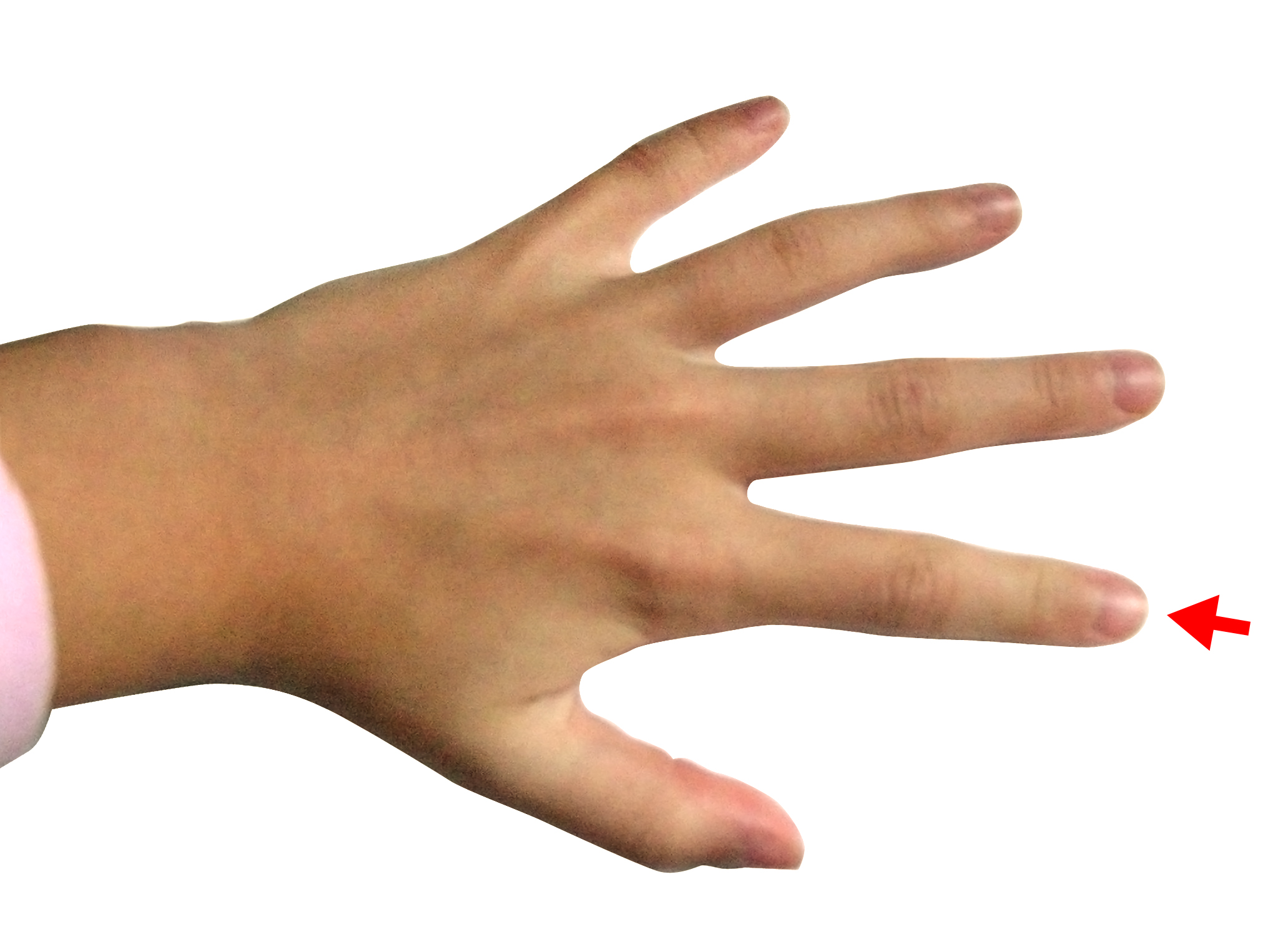
The ratio of index finger length to ring finger length provides an index of sexual differentiation (source: Wikicommons)


Are men and women more alike in some populations than in others? It's possible. First, boys and girls differentiate from each other to varying degrees during adolescence, and this process of sexual differentiation is genetically influenced. There are even conditions, like Swyer syndrome, where an individual is chromosomally male (46, XY) and yet develops externally into a woman.

Second, men and women don't have the same sex roles everywhere. According to a survey of 93 nonindustrial cultures, men were expected to dominate their wives in 67% of them, the sexes were expected to be about equal in 30%, and women were expected to dominate their husbands in 3% (Whyte, 1978). Sex roles differ to varying degrees even among hunter-gatherers, who correspond to the earliest stage of cultural evolution. In the tropics, women provide more food through gathering than men do through hunting. The reverse is true beyond the tropics, where women have few opportunities to gather food in winter (Kelly, 1995, pp. 128-132; Martin, 1974, pp. 16-18).

There has thus been a potential for gene-culture co-evolution. Wherever men and women behave more alike, natural selection will tend to level any innate behavioral differences between them. This can come about in several ways, but a particularly common one is to reduce the sex difference in prenatal hormonal exposure, i.e., the ratio of testosterone to estrogen in the uterine environment of the developing fetus.

We have a "handy" way to measure this prenatal influence. It's called the digit ratio: the length of your index finger divided by the length of your ring finger. The lower your 2nd digit to 4th digit ratio (2D:4D), the more you were exposed to testosterone in the womb and the less to estrogen.

English psychologist John T. Manning has pioneered the use of this digit ratio as a way to measure how prenatal male and female hormones influence various behavioral traits. In a recent study, he looked at how prenatal hormones might influence gender equality in different populations. After measuring the digit ratios of participants from 29 countries, his research team averaged the score for each country and compared it with indices of gender equality: women's share of parliamentary seats; women's participation in the labor force, women's education attainment level; maternal mortality rates; and juvenile pregnancy rates. To ensure comparability, all of the participants were of European descent.

The results?

In short, the more similar the two sexes were in 2D:4D, the more equal were the two sexes in parliamentary and labor force participation. The other variables were not as strongly correlated. (Manning et al., 2014)

In general, women from Northwest Europe have more masculine digit ratios, whereas women from farther east and south have more feminine digit ratios. This geographical trend is more pronounced for the right hand than for the left hand. Since the right-hand digit ratio is associated with social dominance, Northwest Europeans may be less sexually differentiated for that particular trait, as opposed to being less sexually differentiated in general.

Presumably, this isn't a new tendency. Women must have been more socially dominant among Northwest Europeans even before the late 19th century and the earliest movements for women's suffrage. So how far back does the tendency go? To medieval times? To pre-Christian times? It seems to go back at least to medieval times and, as such, forms part of the Western European Marriage Pattern:

The status of women differed immensely by region. In western Europe, later marriage and higher rates of definitive celibacy (the so-called "European marriage pattern") helped to constrain patriarchy at its most extreme level.

[...] In eastern Europe however, the tradition of early and universal marriage (usually of a bride aged 12-15 years, with menarche occurring on average at 14) as well as traditional Slavic patrilocal customs led to a greatly inferior status of women at all levels of society. (Women in the Middle Ages, 2014)

Does this geographic tendency go back to pre-Christian times? There is little consensus on this point, as noted in a study of women in Old Norse society:

The conversion of Iceland raises the problem of the impact of Christianity on the female half of the human race. This, in fact, is one of the most controversial issues in women's history. One point of view argues that Christianity was deeply imbued from the beginning with Jewish and Roman patriarchy, which became intensified by an all-male clergy and resulted in misogyny as the most lasting and profound legacy of Christianity for women. An opposite argument claims that the Christian message was fundamentally a liberating force that included women as well, and although the original radicalism of Jesus on this issue, as on so many others, became diluted with time, women were better off during the Christian period and in Christian countries than they had been before and elsewhere. (Jochen, 1995, p. 2)

Perhaps both arguments are true. As I have argued elsewhere, there may have been a "fruitful encounter" between Christianity and pre-existing behavioral tendencies in Northwest Europe, the result being a significantly different form of Christianity (Frost, 2014).
 

References 

Frost, P. (2014). A fruitful encounter, Evo and Proud, September 26
http://evoandproud.blogspot.ca/2014/09/a-fruitful-encounter.html

Jochens, J. (1995). Women in Old Norse Society, Cornell University Press.

Kelly, R.L. (1995). The Foraging Spectrum. Diversity in Hunter-Gatherer Lifeways, Washington: Smithsonian Institution Press. 

Manning, J.T., B. Fink, and R. Trivers. (2014). Digit ratio (2D:4D) and gender inequalities across nations, Evolutionary Psychology, 12, 757-768.
http://www.epjournal.net/wp-content/uploads/EP1207570768.pdf

Martin, M.K. (1974). The Foraging Adaptation - Uniformity or Diversity? Addison-Wesley Module in Anthropology 56. 

Women of the Middle Ages. (2014). Wikipedia
http://en.wikipedia.org/wiki/Women_in_the_Middle_Ages 

Whyte, M. K. (1978). The status of women in preindustrial societies, Princeton, NJ: Princeton University Press.