Showing posts with label tuberculosis. Show all posts

Monday, February 21, 2022

A natural vaccine?


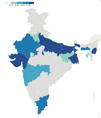
Geographic distribution of the G allele (TIMPRSS2), which is associated with a higher death rate from COVID-19. It’s most frequent on the Indo-Gangetic Plain, which has the longest continuous history of urban settlement in South Asia. Did that environment select for susceptibility to coronaviruses as a way to boost resistance to deadlier respiratory viruses?


The common cold is caused by over 200 strains of rhinoviruses, coronaviruses, adenoviruses, and enteroviruses. Coronaviruses differ from other respiratory viruses in one key respect: they can enter lung tissue via the ACE2 receptor. So if that receptor is altered to allow easier entry, the host would become more susceptible to the common cold but not to other respiratory diseases, including much deadlier ones that cause tuberculosis, pneumonia, or pneumonic plague.


The last point is important because there is evidence that a viral infection can protect against subsequent infection by respiratory viruses. When mice are infected with γherpesvirus 68, which is similar to Epstein-Barr virus, there is production of large quantities of IFN-γ and activation of macrophages that protect against Listeria monocytogenes (which causes listeriosis), Mycobacterium tuberculosis (which causes tuberculosis), and Yersinia pestis (which causes bubonic and pneumonic plague) (Barton et al., 2007; Miller et al., 2019). A cytomegalovirus infection likewise protects against Listeria monocytogenes and Yersinia pestis (Barton et al., 2007).


Coevolution between coronaviruses and early urban settlement


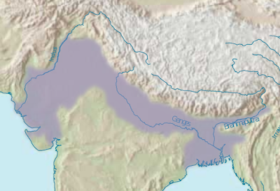
Beginning some 10,000 years ago, hunting and gathering gave way to farming, and nomadism to sedentism. People began to live in progressively larger settlements along the Nile in Egypt, the Tigris and the Euphrates in Mesopotamia, the Indus and the Ganges in northern India, and the Yellow and the Yangtze in China. That is where large numbers of humans first lived in close proximity to each other, and they were particularly vulnerable to the spread of respiratory diseases from one person to another. There may thus have been selection among them for increased susceptibility to coronaviruses, which are normally mild in their effects, as a means to increase resistance to deadlier respiratory viruses.


A recent Indian study by Pandey et al. (2022) suggests that coronavirus susceptibility may have coevolved with risk of infection by life-threatening respiratory viruses like tuberculosis, pneumonia, and pneumonic plague, at least in South Asia. People are more susceptible to infection by coronaviruses if they have the G allele of the TMPRSS2 gene. The research team found that the G allele is significantly associated with a higher fatality rate for COVID-19, apparently because it helps coronaviruses enter lung tissue via the ACE2 receptor.


Pandey et al. (2022) also charted the geographic distribution of the G allele in South Asia. This allele is most frequent among inhabitants of the Indo-Gangetic Plain, i.e., the fertile lowlands that border the Indus and Ganges rivers of northern India and Pakistan. This is also where urbanization has existed for the longest continuous time in South Asia, specifically since the early first millennium BCE. The Indo-Gangetic Plain has had "an uninterrupted sequence of economic development, state formation, and cultural expansion affecting the entire subcontinent as well as Central, East and Southeast Asia" (Heitzman 2008, pp. 12-13).


These findings are roughly consistent with an earlier finding by the same research team. Srivastava et al. (2020) found that an ACE2 allele, at rs2258666, has a negative relationship with the fatality rate for COVID-19. It is also most frequent in the northeast of India, which until recent times was sparsely populated, and whose inhabitants lived in dispersed rural settlements.


References


Barton, E.S., D.W. White, J.S. Cathelyn, K.A. Brett-McClellan, M. Engle, et al. (2007). Herpesvirus latency confers symbiotic protection from bacterial infection. Nature

447: 326-329.

https://doi.org/10.1038/nature05762


Frost, P. (2020). Does a commensal relationship exist between coronaviruses and some human populations? Journal of Molecular Genetics 3(2): 1-2.

https://researchopenworld.com/does-a-commensal-relationship-exist-between-coronaviruses-and-some-human-populations/


Heitzman, J. (2008). The City in South Asia. London: Routledge


Miller, H.E., K.E. Johnson, V.L. Tarakanova, and R.T. Robinson. (2019). γ-herpesvirus latency attenuates Mycobacterium tuberculosis infection in mice. Tuberculosis 116: 56-60.

https://doi.org/10.1016/j.tube.2019.04.022


Pandey, R.K., A. Srivastava, P.P. Singh, and G. Chaubey. (2022). Genetic association of TMPRSS2 rs2070788 polymorphism with COVID-19 case fatality rate among Indian populations. Infection, Genetics and Evolution 98 https://doi.org/10.1016/j.meegid.2022.105206


Shirato, K., M. Kawase, and S. Matsuyama. (2018). Wild-type human coronaviruses prefer cell-surface TMPRSS2 to endosomal cathepsins for cell entry. Virology 517: 9-15.

https://doi.org/10.1016/j.virol.2017.11.012


Srivastava, A., A. Bandopadhyay, D. Das, R.K. Pandey, V. Singh, N. Khanam, N. Srivastava, P.P. Singh, P.K. Dubey, A. Pathak, P. Gupta, N. Rai, G.N.N. Sultana, and G. Chaubey. (2020). Genetic Association of ACE2 rs2285666 Polymorphism with COVID-19 Spatial Distribution in India. Frontiers in Genetics. September 25

https://doi.org/10.3389/fgene.2020.564741


Tuesday, April 7, 2020

COVID-19 update


A dying man, stoned on suspicion of spreading the plague - Felix Jenewein, 1899 (Wikicommons)


SARS-CoV-2, though novel, belongs to a long-existing group of respiratory pathogens: coronaviruses. Until the first appearance of SARS in 2002, these pathogens did little harm to their hosts, usually causing nothing worse than a common cold. So they may have coevolved with us. Furthermore, this coevolution may have taken different forms in different human populations and different cultural environments.

Coronaviruses infect lung tissue via a receptor, ACE2, that varies structurally not only between Asians and other human groups but also between different Asian groups. In particular, the Chinese population has fewer alleles that code for weak binding to the coronavirus S-protein (Cao et al. 2020). Different ACE2 alleles are also associated with differences in susceptibility to diabetic retinopathy, an eye disease with a distinct global pattern of prevalence: 22% in Italy, 23% in China, 30% in the United Kingdom, and 40% in the United States (Adams 2020).

This geographic pattern doesn’t exist because some populations have become more resistant to coronaviruses. Instead, the reverse seems to have happened: some populations have become more susceptible to coronavirus infection, perhaps as a means to prevent more serious pulmonary infections, like tuberculosis and pneumonic plague (Shekhar et al. 2017). Such an effect has been shown with γherpesvirus 68 and cytomegalovirus (Barton et al. 2007; Miller et al. 2019). This crude vaccination boosts the immune response through increased production of IFN-γ and increased activation of macrophages.

Historically, tuberculosis was especially common in crowded environments, where people lived in proximity not only to each other but also to domesticated animals (Comas et al. 2013). Such environments have existed continuously for the longest time in China, as well as in areas like the Indo-Gangetic Plain, the Fertile Crescent, and the Mediterranean Basin. Those areas are where people should be most susceptible to coronavirus infection.

This may explain why COVID-19 has been more severe in southern Europe than in northern Europe. It is surprising that infection tends to become less severe with latitude when one would expect the opposite: respiratory viruses spread more effectively under conditions of lower temperature, lower humidity, and lower solar UV.


Ongoing research?

These geographic differences have caught the interest of a molecular epidemiologist at the University of Hawai'i, Maarit Tiirikainen:

"There have been major differences in the rates of SARS-CoV-2 infection and the severe disease between the different geographic regions since the beginning of the COVID-19 pandemic, even among young individuals," Dr. Tiirikainen said. "Epidemiological studies-so-called Genome Wide Association Studies (GWAS)-indicate that populations carry different variants of the ACE2 gene. This variation in the gene coding for the ACE2 receptor may have an effect on the number of ACE2 receptors on the lung cells, as well as on how effectively the virus binds to the receptor. There may also be genetic differences in immune and other important genes explaining why some people get more sick than others."

She is collaborating with a genomics company, LifeDNA, in a study that will initially focus on Hawai'i's multiethnic inhabitants, specifically their diversity of ACE2 alleles in relation to the latest coronavirus (LifeDNA 2020 – h/t to Steve Sailer).


Parting thoughts

All humans can get infected by coronaviruses, but the infection tends to vary in severity from one population to another. This variance may reflect differences in genetic adaptation in different cultural environments.

Of course, adaptation may also be cultural. Because natural selection acts on the end result, and not on the means to that end, the means may be a purely learned algorithm, like adding spices to food or avoiding physical contact with strangers. One might not have understood why or how such practices worked, but they did work and would be passed on to subsequent generations, thus becoming the traditional way of doing things. Today, we’re likely to reject such practices as outmoded superstitions.

So be modern. Hug a stranger.


References

Adams N. (2020). Cracking the code to the 2019 novel coronavirus (COVID-19): Lessons from the eye. Eye Reports 6(1). 
https://eyereports.org/index.php/eyereports/article/view/97

Barton E.S., White D.W., Cathelyn J.S., Brett-McClellan K.A., Engle M., Diamond M.S., et al. (2007). Herpesvirus latency confers symbiotic protection from bacterial infection. Nature 447: 326-329. 
https://www.nature.com/articles/nature05762

Cao Y., Li L., Feng Z., Wan S., Huang P., Sun X., et al. (2020). Comparative genetic analysis of the novel coronavirus (2019-nCoV/SARS-CoV-2) receptor ACE2 in different populations. Cell Discovery 6(11). 
https://www.nature.com/articles/s41421-020-0147-1%3C/blockquote%3E 

Comas I., Coscolla M., Luo T., Borrell S., Holt K.E., Kato-Maeda M., et al. (2013). Out-of-Africa migration and Neolithic coexpansion of Mycobacterium tuberculosis with modern humans. Nature Genetics 45(10): 1176-1182.
https://www.ncbi.nlm.nih.gov/pmc/articles/PMC3800747/

LifeDNA (2020). COVID-19: LifeDNA and University of Hawai’i Collaborate on Studying Why Certain Populations Are Hit Harder. Research focuses on ACE2 receptor, probing the role of genetics in both susceptibility to infection and severity of response April 2, University of Hawai'i Cancer Center 
https://www.uhcancercenter.org/about-us/newsroom/600-covid-19-lifedna-and-university-of-hawai-i-collaborate-on-studying-why-certain-populations-are-hit-harder

Miller H.E., Johnson K.E., Tarakanova V.L., Robinson R.T. (2019). γ-herpesvirus latency attenuates Mycobacterium tuberculosis infection in mice. Tuberculosis 116: 56-60. 
https://www.ncbi.nlm.nih.gov/pmc/articles/PMC6876742/

Shekhar S., Schenck K., Petersen F.C. (2017). Exploring host-commensal interactions in the respiratory tract. Frontiers in Immunology 8: 1971. 
https://www.frontiersin.org/articles/10.3389/fimmu.2017.01971/full

Monday, March 2, 2020

Coevolution with the plague


Houses being burned during an outbreak of pneumonic plague in China (Wikicommons). In China, large urban populations coevolved with deadly pulmonary infections, like tuberculosis, pneumonia, and pneumonic plague. Because resistance was boosted by regular exposure to normally mild infections by coronaviruses, there was natural selection for more susceptibility to them.


Two posts ago I argued that coronaviruses have coevolved with the Chinese population, to the point of developing a commensal relationship. A few points:

-  Such viruses include the common cold and are normally mild in their effects.

- Repeated coronaviral infections of lung tissue may actually help increase resistance to more serious pulmonary infections, like tuberculosis, pneumonia, and the Spanish flu of 1918—which curiously spared China. 

- Chinese lung tissue would thus facilitate coronaviral infections as a sort of routine vaccination. 

- If this is true, modern medicine has inadvertently made the Chinese population particularly vulnerable to deadly diseases like the Wuhan coronavirus by reducing the prevalence of milder pulmonary infections.


The examples of herpesvirus and cytomegalovirus

This cross-immunity is seen with other viruses. In mice, gammaherpesvirus 68 (similar to Epstein-Barr virus) provides immunity against much deadlier bacterial pathogens: Listeria monocytogenes; Yersinia pestis, which has caused plagues like the Black Death; and Mycobacterium tuberculosis, i.e. tuberculosis (Barton et al. 2007; Miller et al. 2019). Infection with cytomegalovirus likewise immunizes against Listeria monocytogenes and Yersinia pestis (Barton et al. 2007).

Quite a few writers have argued that many common pathogens are actually allies that help us fight more serious diseases: 

The microbial communities of humans are characteristic and complex mixtures of microorganisms that have co-evolved with their human hosts. The species that make up these communities vary between hosts as a result of restricted migration of microorganisms between hosts and strong ecological interactions within hosts, as well as host variability in terms of diet, genotype and colonization history. The shared evolutionary fate of humans and their symbiotic bacteria has selected for mutualistic interactions that are essential for human health, and ecological or genetic changes that uncouple this shared fate can result in disease. (Dethlefsen et al. 2007)

So it’s possible that humans have coevolved with mildly acting viruses as a means to ward off pathogens that cause more serious pulmonary infections, such as pneumonic plague and tuberculosis. Moreover, this coevolution may have taken different forms in different human populations, a possibility raised by Miller et al. (2019): "our results suggest human γHV-infection may be an important but unrecognized factor which modifies TB outcome, particularly in high TB burden countries where most children acquire EBV [Epstein-Barr virus] by 3 years of age."

Comas et al. (2013) describe the evolution of tuberculosis in our species and how it became more common in certain human environments, particularly "crowded" ones:

Crowd diseases are generally highly virulent and depend on high host population densities to maximize pathogen transmission and reduce the risk of pathogen extinction through exhaustion of susceptible hosts. Many crowd diseases emerged during the Neolithic Demographic Transition (NDT) starting around ten thousand years ago (kya), as the development of animal domestication increased the likelihood of zoonotic transfer of novel pathogens to humans, and agricultural innovations supported increased population densities that helped sustain the infectious cycle. The marked expansion of MTBC [Mycobacterium tuberculosis complex] during the NTD, but not during earlier human expansion events, suggests that the success of this pathogen was primarily driven by increases in human host density, which is typical of crowd diseases.

Perry et al. (2010) have shown that Helicobacter pylori, a bacterium that lives in the stomach lining, greatly reduces the risk of tuberculosis infection. Again, less serious infections help ward off much more serious ones, like tuberculosis:

Why only 10% of infected individuals succumb to tuberculosis remains one of the most vexing public health questions—one which the one-pathogen-one-disease paradigm is ill-equipped to answer. While preliminary, our work suggests that one factor contributing to the clinical outcome of TB infection may be a concurrent chronic infection. The hypothesis that the human microbiome has evolved to provide context-specific competitive risk advantages to the host also raises the intriguing possibility that our microbiota can be manipulated to modulate disease risk from M. tuberculosis, as well as other common human pathogens. (Perry et al. 2010)


Have the Chinese coevolved with coronaviruses?

Common viral infections may have a similar protective effect. If we go back to the Barton et al. study, we find that it was criticized by Yager et al. (2009) on the grounds that the cross-immunity seems to last only five months after acute infection. To benefit from this cross-immunity, lung tissue should therefore be regularly infected with a virus whose adverse effects are both mild and temporary, like most coronaviruses.

Are the Chinese innately more susceptible to coronaviruses? Attention has focused on a study by Zhao et al. (2020), who, using lung tissue from several donors, studied a receptor, ACE2, that acts as the point of entry for some coronaviruses, including the one responsible for the outbreak in Wuhan. They found that the receptor was concentrated in certain cells and that the number of such cells in lung tissue was five times greater in the Asian donor. Yes, there was only one Chinese donor, but the chances are very low that the same normal distribution would produce such an extreme outlier.

This finding is also consistent with those of previous studies. Cheng et al. (2007) looked at other receptors for viral infections and found differences between Chinese and other human populations. In the specific case of pulmonary diseases, Seitz et al. (2012) studied the prevalence of bronchiectasis in the United States and found a prevalence 2.5 to 3.9 times higher among Asian Americans than among Euro Americans or African Americans. Kwak et al. (2010) likewise found a high prevalence of bronchiectasis in Korean adults.

Since my last post on the subject, two more studies have come out.


The Cai study

Cai (2020) failed to find significant differences in ACE2 receptor gene expression between Asian and Caucasian lung tissue but did find an interaction between smoking history and ethnicity: "we found ACE2 is most actively expressed in AT2-reformed cells in former Asian smokers but not in Caucasian current smokers and African American non-smokers." However, this difference wasn’t significant. 

This study has an adequate sample size (n=345) but uses a questionable classification by ethnicity. The lung tissue samples were from a U.S. company, Gene Expression Omnibus, which classifies its samples as "Caucasian," "African American," or "Asian." Although most Asian Americans are of East Asian descent, many have roots in Southeast Asia or South Asia. As we will see, there are probably significant differences in the ACE2 receptor even between Asian groups.


The Cao et al. study 

Cao et al. (2020) looked at the different alleles for the ACE2 receptor gene in two databases: the China Metabolic Analytics Project and the 1000 Genomes Project. They found large differences in allele frequencies among human populations, not only between Asians and other human groups but also between different Asian groups. "These data suggested that there was a lack of natural resistant mutations for coronavirus S-protein binding in [some] populations."

Their conclusion more or less sums up current knowledge:

Recent reports of the ACE2 expression analysis in lung tissues from Asian and Caucasian populations are still controversial. The single-cell RNA-seq analysis reported that the Asian donor had much higher ACE2 expression cell ratio than white and African-American donors. In contrast, the ACE2 expression analysis using the RNA-seq and microarray datasets from control lung tissues indicated there were no significant differences between Asian and Caucasian, or male and female. The ACE2-expressing cells are a very small part of cells in lung tissues. The sample size and the purity of ACE2-positive cells in the selected samples would influence the conclusions. Our analysis showed the differences in distribution and AFs [allele frequencies] of eQTLs for ACE2 in different populations, indicating the diversity of ACE2 expression pattern in populations. […] In addition, our data showed the moderate difference in AFs of eQTLs between South Asian and EAS [East Asians], which suggests the potential difference of ACE2 expression in different populations and ethnics in Asia. (Cao et al. 2020)


Conclusion

Without knowing what these alleles actually do, we can only say that the ACE2 receptor has coevolved differently with different human populations and, presumably, different natural and social environments. In particular, crowded environments, with high rates of life-threatening pulmonary infections, notably tuberculosis, pneumonia, and pneumonic plague, should have favored individuals who are more susceptible to infection by coronaviruses. 

Historically, such environments would encompass not only China but also other areas that have long had large urban populations and a correspondingly long coevolution with pulmonary infections. These areas would notably include the Indo-Gangetic Plain in India and the Fertile Crescent of the Middle East.


References

Barton, E.S., D.W. White, J.S. Cathelyn, K.A. Brett-McClellan, M. Engle, M.S. Diamond, V.L. Miller. H.W. Virgin IV. (2007). Herpesvirus latency confers symbiotic protection from bacterial infection. Nature 447:326-9.
https://www.nature.com/articles/nature05762

Cai, G. (2020).  Bulk and single-cell transcriptomics identify tobacco-use disparity in lung gene expression of ACE2, the receptor of 2019-nCov. medRxiv February 17
https://www.medrxiv.org/content/10.1101/2020.02.05.20020107v2

Cao, Y., L. Li, Z. Feng, et al. (2020). Comparative genetic analysis of the novel coronavirus (2019-nCoV/SARS-CoV-2) receptor ACE2 in different populations. Cell Discovery 6: 11.
https://www.nature.com/articles/s41421-020-0147-1

Cheng, P-L, H-L. Eng, M-H. Chou, H-L. You, T-M. Lin, (2007). Genetic polymorphisms of viral infection-associated Toll-like receptors in Chinese population. Translational Research 150(5): 311-318
https://www.sciencedirect.com/science/article/pii/S1931524407000953

Comas, I., M. Coscolla, T. Luo, S. Borrell, K.E. Holt, M. Kato-Maeda, et al. (2013). Out-of-Africa migration and Neolithic coexpansion of Mycobacterium tuberculosis with modern humans. Nature genetics 45(10): 1176-1182.
https://www.ncbi.nlm.nih.gov/pmc/articles/PMC3800747/

Dethlefsen, L., M. McFall-Ngai, and D. Relman. (2007). An ecological and evolutionary perspective on human-microbe mutualism and disease. Nature 449: 811-818.
https://www.researchgate.net/profile/David_Relman/publication/5902740_Dethlefsen_L_McFall-Ngai_M_Relman_DA_An_ecological_and_evolutionary_perspective_on_human-microbe_mutualism_and_disease_Nature_449_811-818/links/0deec5278790b8a5be000000.pdf

Kwak, H.J., J.Y. Moon, Y.W. Choi, T.H. Kim, J.W. Sohn, H.J. Yoon, D.H. Shin, S.S. Park, and S.H. Kim. (2010). High prevalence of bronchiectasis in adults: analysis of CT findings in a health screening program. Tohoku Journal of Experimental Medicine 222: 237-242.
https://pdfs.semanticscholar.org/dd5d/c5d64f82c84277b74024af0671c8ec070fa6.pdf  

Miller, H. E., K.E. Johnson, V.L. Tarakanova, and R.T. Robinson. (2019). γ-herpesvirus latency attenuates Mycobacterium tuberculosis infection in mice. Tuberculosis (Edinburgh, Scotland) 116: 56-60.
https://www.ncbi.nlm.nih.gov/pmc/articles/PMC6876742/

Perry, S., B.C. de Jong, J.V. Solnick, M. de la Luz Sanchez, S. Yang, P.L. Lin, et al. (2010). Infection with Helicobacter pylori is associated with protection against tuberculosis. PloS one 5(1), e8804.
https://www.ncbi.nlm.nih.gov/pmc/articles/PMC2808360/

Seitz, A.E., K.N. Olivier, J. Adjemian, S.M. Holland, and D.R. Prevots. (2012). Trends in bronchiectasis among medicare beneficiaries in the United States, 2000 to 2007. Chest 142(2): 432-439.
https://www.ncbi.nlm.nih.gov/pmc/articles/PMC3425339/  

Yager, E.J., F.M. Szaba, L.W. Kummer, K.G. Lanzer, C.E. Burkum, S.T. Smiley, and M.A. Blackman. (2009). γ-Herpesvirus-induced protection against bacterial infection is transient. Viral immunology 22(1): 67-72.
https://www.ncbi.nlm.nih.gov/pmc/articles/PMC2952138/

Zhao, Y., Z. Zhao, Y. Wang, Y. Zhou, Y. Ma, and W. Zuo. (2020). Single-cell RNA expression profiling of ACE2, the putative receptor of Wuhan 2019-nCov. bioRxiv January 26
https://www.biorxiv.org/content/10.1101/2020.01.26.919985v1.full  


Thursday, July 16, 2009

Who gets to write history?

History is written by the survivors – Max Lerner

I prefer Lerner’s version to Churchill’s History is written by the victors. Opinions will survive as long as the group that holds them, and such a group may disappear for reasons besides defeat through conflict. Often, the group is temporary by nature.

Remember this when reading about how the sunshine movement eradicated rickets and tuberculosis—the two long-running epidemics of the Western world. This is a history written by those who kept making their case long after others had lost interest. To quote a Sailerism: History is written by those who write … and write … and write.

Here is how the sunshine movement is usually presented:

In the nineteenth century, different physicians advocated ‘heliotherapy’ as a means to treat rickets and kill the microbes that cause tuberculosis. They were ignored, except by a few nudists and health faddists. Then, two events broke the barrier to public acceptance. One was the Spanish flu of 1918-1919, which showed the need for strong measures to keep microbes from spreading. The other, in 1919, was the discovery that ultraviolet light can cure rickets by releasing a chemical into the bloodstream, later identified as vitamin D. Heliotherapy thus gained in popularity and by the late 1920s sunbathing had become widespread. Meanwhile, tuberculosis and rickets steadily declined until both virtually disappeared.

This narrative leaves out a key detail. The sunshine movement had little effect on the prevalence of either tuberculosis or rickets. Through the 1920s and 1930s, there was only a gradual decline in the incidence of tuberculosis, as part of a downward trend that began in the nineteenth century and that probably was due to stricter segregation of tubercular patients in hospitals and sanatoria. Steep decline began later—after the Second World War (
Wilson, 1990). Much of the credit goes to the discovery of antimicrobial drugs like isoniazid, rifampicin, and streptomycin. Just as important was the overall rise in the standard of living, particularly among urban workers. As the population became better nourished and as overcrowded tenements gave way to suburban homes, microbes could not spread so easily to weakened hosts.

Nor was rickets much affected. In Dundee, Scotland, its incidence among children held steady from 1925 to 1935 and then dropped abruptly, apparently after the introduction of a ‘milk in schools’ scheme in 1934 and then greater provision of free meals to needy children. This drop preceded vitamin D fortification of milk and infant cereals (Stewart et al., 1964). In the United States, rickets was deemed as late as 1940 to be “still probably the most common disease of early childhood” (Harrison, 1966). In Sweden, it still afflicted most fetuses and newborns during the early 1950s (Sydow et al., 1956).

By this time, many medical researchers had concluded that the cause could not be lack of sun. A Swedish autopsy study found that rickets in fetuses and newborns showed no significant correlation with the mother’s vitamin D status during pregnancy, as indicated by her consumption of vitamin D supplements or by her degree of sun exposure, i.e., daily walks in the sun, outdoor work, summer holiday in the country, and latitude within Sweden (Sydow et al., 1956). For several research teams, the cause seemed to be substances in commercial bread, like phytic acid, that immobilize calcium and phosphorus within the body (Bruce & Callow, 1934; Harrison & Mellanby, 1939; McCance et al., 1942; McCance & Widdowson, 1942). But fewer and fewer researchers were now interested. Like tuberculosis, rickets too went into a steep decline after the war, eventually becoming a medical oddity by the 1960s (Harrison, 1966).

Meanwhile, interest had been growing in the sunshine movement. It now extended far beyond the medical community, being influential in the fashion industry, the arts and literature, architecture, and the movie industry. It had become a cultural phenomenon, and one that would create much of the look and feel of modern life.

Fashion industry

Gabrielle (Coco) Chanel is usually credited with making suntans popular among women. Certainly, in the late 1920s she was the one who added tanned skin to a new androgynous look, la garçonne, that featured long legs, a flat chest, narrow hips, and large shoulders, like a young boy on the brink of puberty (Andrieu, 2008, p. 73; Bard, 1998; Galante, 1972; Wilson, 1985).

But Coco Chanel simply boarded a bus that was already on the road. As early as 1926, a Connecticut radio station announced: “a coat of tan seems to be the latest style in natural coloring at this season of the year. [It has] been increasing in favor during the last few years” (Nickerson, 1926). Female tanning became fashionable through women acting on their own initiative. Others then saw the market opportunities:

Cosmetics manufacturers took notice of the new acceptability of nonwhite skin and began to produce darker powders, as well as artificial bronzing lotions. By 1929, Jean Patou and Coco Chanel had introduced suntan products, and Helena Rubenstein was selling “Valaze Gypsy Tan Foundation.” Other cosmetics manufacturers were blending powders to be “creamy,” rather than white, and producing “ochre,” “dark rachel,” and “suntan” shades. (Berry, 2000, p. 188)

During this time, skin whiteners became less popular.

Golden Peacock Bleach Cream and other facial bleaches, which were advertised regularly in women’s magazines until the late 1920s, appeared only rarely after the early 1930s, although skin lighteners were still marketed to the African American community. (Berry, 2000, p. 188)

The same trend swept through women’s magazines. By the end of the 1920s, Vogue was telling its readers that “The 1929 girl must be tanned” and “A golden tan is the index of chic” (Vogue, 1929). In the early 1930s, however, these magazines were periodically predicting the end of the suntan fad (Berry, 2000, p. 188). Just as fashion leaders had failed to anticipate this fad, they also misjudged its staying power.

Arts and literature

In the mid-1920s, the sunshine movement spread to artists and literati through multiple points of entry. A key one was the French Riviera (Ash, 1974; Weigtman, 1970).


Among the fashionable, “heliophobia” soon gave way to “heliophilia.” The scene of this minor revolution was the French Riviera—specifically the beach of La Garoupe at Cap d’Antibes—and its chief ideologist was Gerald Murphy [an ex-pat American artist].

… It was the Americans—Cole Porter and the Murphys—who first “discovered” the Riviera as a summer resort. The Murphys began to clear La Garoupe of its layer of seaweed and persuaded the proprietor of the Hotel du Cap to remain open during the summer months … The cultivators of the simple, the connoisseurs of the primitive, formed a new elite comprised of Americans, artists and the more unconventional members of the aristocracy. Among others, the Hemingways, the Fitzgeralds, the Picassos, the Legers and the Count and Countess Etienne de Beaumont, all joined the Murphys at Cap d’Antibes in the summer. It was this elite, with some assistance from Coco Chanel, that raised the suntan to the level of higher fashion. (Ash, 1974)

Andrieu (2008, p. 73) and Weigtman (1970) describe how French writers of the 1920s placed favorable references to tanned skin in their works, associating it with lead characters and positive qualities. Similar placement appears in F. Scott Fitzgerald’s The Great Gatsby (1925). Miss Jordan Baker has "sun-strained eyes," a "slender golden arm," a "brown hand," a "golden shoulder," and a "face the same brown tint as the fingerless glove on her knee” (Fitzgerald, 1992, pp. 15, 47, 57, 84, 185).

Architecture

The sunshine movement brought a new urban landscape by moving buildings further back from the street, limiting their height, and spacing them further apart. Windows also became bigger and more numerous. Meanwhile, modernist architects looked to tuberculosis sanatoria and ocean liners to make their creations more open to the sun and air. They introduced such features as the flat roof, the balcony, and the roof or garden terrace “on which elegant Jazz age young women, dressed in patio-pyjamas, could sunbathe on chaises longues” (Campbell, 2005).

These signatures of modernism marked the design of public housing and, especially, schools, whose population was thought to be most at risk for tuberculosis and rickets:

By the late 1920s, the therapeutic qualities of sunlight were widely recognised, and its use was extended in sunshine schools and open-air clinics to the more general treatment of sickly, TB-prone and crippled children, many of them drawn from the slums. Progressive schools like Bedales early encouraged sunbathing; St Christopher School, Letchworth, installed vita glass; and pictures of Pinehurst School show the children running about naked. (Twigg, 1981)

In general, traditional architecture was seen not simply as old-fashioned but as unhealthy. This gave a revolutionary urgency to the thinking of modernists, like the Swiss architect Le Corbusier:

But when it comes to a question of demolishing rotten old houses full of tuberculosis and demoralizing, you hear them cry, “What about the iron-work, what about the beautiful old wrought-iron work.” (Campbell, 2005)

Such demolition was considered necessary to build a healthier society. “Sunlight stood for the new society of light”:

Houses in the garden cities were oriented towards the sun. Architecture in the interwar years pursued light to an almost obsessive degree. It came to be the emblem of a cluster of reforms in the 1920s aimed at making Britain a better, healthier, cleaner place to live.

… Just as the antiseptic qualities of sunlight had been observed through its action on mouldy, damp objects, so the sunlight for this post-Victorian generation could be made to shine on the dank, rotten and hidden aspects of the Victorian world. (The thirties saw the full flood of anti-Victorianism) This could mean the slum houses and sick children, but it also, very frequently, meant sexuality.
(Twigg, 1981)

Movie industry

The tanned look entered the movie industry via individual actors and actresses, notably Joan Crawford:


Joan Crawford was credited for spreading the trend among Hollywood flappers—in addition to tanning her face, Crawford browned her body and went stockingless …
(Berry, 2000, p. 188)


She was reportedly told by MGM to stop tanning because it made her look “like a lineal descendent of Sheba.” The movie industry, however, soon realized there was a market for dark skin as an item of sexual interest. The 1930s thus saw a spate of Hollywood films featuring Latin lovers, Arab sheikhs, and South Seas beauties (Berry, 2000, pp. 110-111).

No one fully understood this phenomenon. Described as a quest for the exotic, one might doubt the exoticism of stars and starlets who were just a darker version of the European phenotype. Nonetheless, through productions like South Pacific (1949), this faux interracialism would help pave the way for the real thing after the war.

Perhaps white Americans were motivated to sexualize dark skin by a ‘rare color effect’ such as exists with differing shades of hair color. The rarer brunettes are, the more they excite sexual interest in men (
Anon, 2008; Thelen, 1983). Or perhaps the motivation lay at another level of male sexual response. If women evolved a lighter complexion and other paedomorphic features as a way to inhibit male aggression and stimulate feelings of care, a darker skin tone could exert sex appeal on a more aggressive and less empathetic level (Frost, 2007; Guthrie, 1970).

Social conformity and status competition

Cultural change involves not only leaders but also followers. As more and more people sported tans, the new look tended to spread simply through social conformity and status competition:

The appearance of medical articles that begin to deal with tanning as not directly related to a “cure” shows that the acquisition of the suntan, at least among certain sections of society, was already desirable. In addition the change toward a more positive view of the suntan was to articulate well with other social changes taking place, particularly as related to travel, in the early twentieth century. The idea of health travel was established, with many invalids seeking the sun cure in sanatoria. However, the wealthy invalid was able to travel further — to locations where they could be assured of receiving sunlight, such as the Swiss Alps or the Mediterranean. (Carter & Michael, 2003, p. 269)


The cosmetic aspects of a suntan were not originally much to the fore; during the 1920s tanning was regarded as only a side effect and not spoken of with special favour. By the 1930s, however, the naturist magazines were praising the look of bronzed skin. The fashion spread beyond these circles, to the cosmopolitan and wealthy. By the 1930s the Riviera season had reversed from being winter to summer. The seaside, from being a place for bathing and for sea air became somewhere for taking off your clothes and lying in the sun; the resorts began to publish their sunshine figures; and by the mid 1930s the major cosmetic houses were producing suntan creams. A suntan became associated with youth, health and vigour, qualities that the thirties found particularly attractive sexually. (Twigg, 1981)

During the 1930s, the popularity of suntanning and nudity reached a peak, intellectual and social benefits were said to accrue from sun-exposure, and it was felt to be “imperative” for the successful executive to be tanned, as this indicated “superior physique, intelligence, and moral character.” (Koblenzer, 1998)


Conclusion

The sunshine movement was able to impose its world view—and its version of history—by winning over the creators of modern culture: artists, writers, actors, architects, and fashion designers. This victory, if we can use the term, was achieved not in the narrow realm of medical debate but in the larger one of cultural production. In truth, there was no victory because there was no battle. The ‘other side’ lost interest in explaining the tuberculosis and rickets epidemics once these had subsided. They moved on to other things.

The ‘victors’ had an unforeseen ally: a sensual, if not sexual, fascination with dark skin. By arming young men and women with a medical alibi, the sunshine movement unwittingly opened up a dimension of sexual attraction that had lain unexploited. There had, in fact, been a taboo against sexualizing dark skin, partly because of the racial connotations and partly because dark complexions among white Americans had traditionally been viewed as unfeminine for women and as hypermasculine for men.

Another ally, widespread in the twentieth century, was a belief in change and in the urgency of change. It is probably no coincidence that many sunshine advocates, like the architect Le Corbusier, saw themselves as radicals. The movement likewise had more success in pushing its agenda in the Eastern bloc, as seen in the mass relocation of working families to modernist housing projects, in State-sponsored vacations at Black Sea resorts, and in the DDR’s mass administration of vitamin D megadoses to children.

References

Andrieu, B. (2008). Bronzage. Une petite histoire du Soleil et de la peau, CNRS Éditions.

Anon. (2008).
Maxim's audience prefers brunettes; distribution is bimodal. Gene Expression, July 6, 2008.

Ash, J. (1974). The meaning of suntan, New Society, August, pp. 278-280.

Bard, C. (1998). Les garçonnes. Modes et fantasmes des Années folles. Paris: Flammarion.

Berry, S. (2000). Screen Style. Fashion and Femininity in 1930s Hollywood, Minneapolis: University of Minnesota Press.

Bruce, H. & Callow, R. (1934). Cereals and rickets. The role of inositolhexaphosphoric acid, Biochemical Journal, 28, 517-528.

Campbell, M. (2005). What tuberculosis did for modernism: The influence of a curative environment on modernist design and architecture, Medical History, 49, 463-488.

Carter, S. & Michael, M. (2003). “Here comes the sun: Shedding light on the cultural body”, in: H. Thomas & J. Ahmed (ed.) Cultural Bodies: Ethnography and Theory, pp. 260-282, Wiley-Blackwell.

Fitzgerald, F.S. 1992. The Great Gatsby. New York: Collier Books.

Frost, P. (2007). Comment on Human skin-color sexual dimorphism: A test of the sexual selection hypothesis, American Journal of Physical Anthropology, 133, 779-781.

Galante, P. (1972). Les années Chanel. Paris: Mercure de France.

Guthrie, R.D. (1970). Evolution of human threat display organs, Evolutionary Biology, 4, 257-302.

Harrison, D.C., & Mellanby, E. (1939). Phytic acid and the rickets-producing action of cereals, Biochemical Journal, 33, 1660-1680.

Harrison, H.E. (1966). The disappearance of rickets, American Journal of Public Health, 56, 734-737.

Koblenzer, C.C. (1998). The psychology of sun-exposure and tanning, Clinics in Dermatology, 16, 421-428.

McCance, R., & Widdowson, E. (1942). Mineral metabolism of dephytinized bread, Journal of Physiology, 101, 304-313.

McCance, R., & Widdowson, E. (1942). Mineral metabolism of healthy adults on white and brown bread dietaries, Journal of Physiology, 101, 44-85.

Nickerson, E.C. (1926). Nature's Cosmetics, Bulletin sanitaire 26(5), 134-140.

Stewart, W.K., Mitchell, R.G., Morgan, H.G., Lowe, K.G., & Thomson, J. (1964). The changing incidence of rickets and infantile hypercalcaemia as seen in Dundee, The Lancet, 283(7335), 679-730.

Sydow, G.V., Ranström, S., Berezin, D., & Axen, O. (1956). Histological findings characteristic of rickets in foetuses and young infants, Acta Paediatrica, 45, 114-138.

Thelen, T.H. (1983). Minority type human mate preference. Social Biology, 30, 162-180.

Twigg, J. (1981).
“Sunlight & nature” in: The Vegetarian Movement in England, 1847-1981: A study in the structure of its ideology, Doctoral thesis presented to the London School of Economics, University of London. http://www.ivu.org/history/thesis/sunlight.html

Vogue (1929). June 22, pp. 99, 100.

Weightman, J. (1970). The solar revolution. Reflections on a theme in French literature, Encounter, December, pp. 9-18.

Wilson, E. (1985). Adorned in Dreams: Fashion and Modernity, London: Virago.

Wilson, L.G. (1990).
The historical decline of tuberculosis in Europe and America: Its causes and significance, Journal of the History of Medicine and Allied Sciences, 45, 366-396.